VueBox® Product information
VueBox® is a quantification software designed to analyze DCE-US DICOM clips and is intended for Clinicians and Researchers interested in:
- Quantification of DCE-US sequences
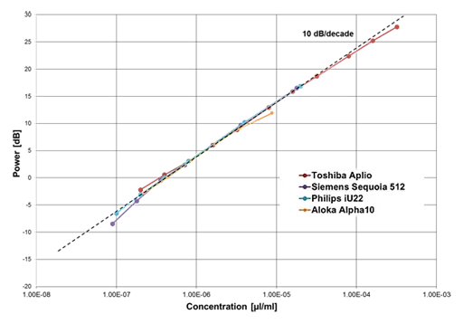
- Excellent cross-platform reproducibility due to dedicated calibration files for all major ultrasound scanners
- Obtaining reliable perfusion parameter values
- Documenting analyses in a synthetic report
- Retrieving and comparing examinations performed at different dates
- Documenting their work for publication purposes
- Improving interobserver agreement
What is quantification?
Perfusion is a recognized parameter of tissue functionality and vitality, providing the clinician with a critical insight into the health of the tissue. Quantification of blood volume and flow is crucial for determining areas of different perfusion in whole organs/tissues due to impairment of blood supply or to characterize different lesions on the anatomical and functional aspects of vascular physiology.
What is the software?
VueBox® is a general-purpose software application for quantifying tissue perfusion using Dynamic Contrast-Enhanced Ultrasound (DCE-US):
VueBox® is a quantification toolbox designed to analyze DCE-US DICOM clips obtained with a wide range of ultrasound systems. Its unique Bracco-patented technology and linearization process allow quantitative assessment of perfusion.
Key features:
- Solution to analyze DICOM clips from different ultrasound systems
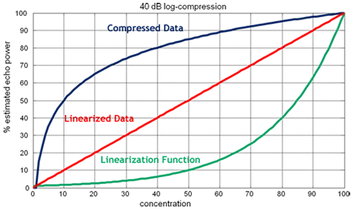
- Linearization of video data for accurate measurements
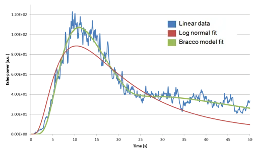
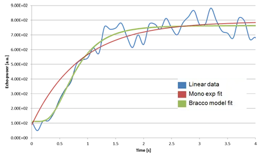
- Optimized curve fitting based on Bracco-patented technology
- Compatible with bolus and replenishment kinetics
- Multiple parametric images
- Short processing time thanks to parallel programming optimized for multi-core processors
Additional features:
- Fully automatic motion compensation
- Easy-to-use clip editor
- Multiple window interface using tabs
- Concatenation of multiple clips
- Automatic detection of contrast arrival
- Saving and retrieving of user-drawn Regions of Interest
- Automatic management of side-by-side display(contrast and B-mode)
- Length and area measurements
- Real time clip player
- Clip anonymization
Management of analysis results:
- Saving and retrieving of analysis results and context
- Export of graphs and images (BMP, TIF, JPEG),data (Excel compatible) and clips (WMV)
- Generator of customizable and easy-to-read analysis report
Who will benefit of the software?
VueBox® is intended for Clinicians and Researchers interested in:
- DCE-US perfusion quantification in any type of tissue
- Obtaining reliable perfusion parameter values
- Processing data acquired with different ultrasound platforms
- Documenting analyses in a synthetic report
- Retrieving and comparing examinations performed at different dates
- Documenting their work for publication purposes
| Brochure | 04/20/2023 | Download |
-
GI-Perfusion
-
To quantify blood perfusion in organs (non-cardiac indications)
Provide a reliable method for quantifying organ perfusion, obtained from DCE-US examinations; -
With specific tools to follow-up perfusion of the same tissue or lesion over time
Provide means for documenting and comparing analyses performed at different patient examinations (visits).
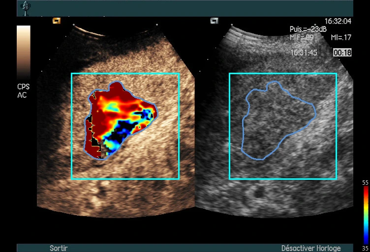
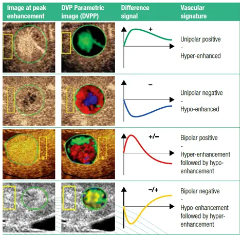
Liver DVP
-
To highlight differences in contrast agent perfusion kinetics in focal liver lesions
Provide tools to summarize the specificities of the difference signals signatures into a single, easy-to-read color-coded image.
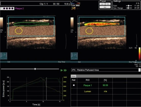
Plaque package
-
to detect vascularization and determine vascularized areas in atherosclerotic plaques
-
specific vascularization parameters : plaque area, perfused area, relative perfused area, mean opacification.
-